
This is a 9 year old girl who presents with wheezing for the first time in the setting of fever. You can see below between the two rib shadows the beginnings of a consolidated lung with air bronchograms (which shows up as white) consistent with a pneumonia. You also see that the A-lines are obliterated, […]
Author: Carrie Ng
Carrie was born in San Francisco, but grew up between Hong Kong and Los Angeles. Although she loves the sun, she was excited to move away from home to Philadelphia for medical school, where she explored a new city, learned to appreciate all four seasons, and met her Canadian husband. She then moved to New York City and completed her Pediatrics residency at NYU and PEM fellowship at Columbia. During this time, Carrie developed a passion for bedside ultrasound and decided to join the CNMC faculty while pursuing an Emergency Ultrasound fellowship. In her spare time, Carrie loves traveling, listening to podcasts, and playing board games.
IOTW: Pilodinal cyst with echogenic fat
Pt is a teen with a clinical presentation of a pilonidal cyst with abscess. It’s interesting because you see the very bright structures in the far field: that’s echogenic fat that can often clump in these collections. Often we think of hyperechoic structures being bone, but these are not teeth for example since they don’t shadow as […]
IOTW: WES sign
This is a 16 y/o F w/ RUQ pain and scleral icterus. This week’s image shows the WES sign which stands for Wall-Echo-Shadow. As you can see, sometimes the gallbladder is contracted around a large gallstone and all you see is the gallbladder wall, hyperechoic gallstone followed by shadowing behind the stone, preventing you from […]
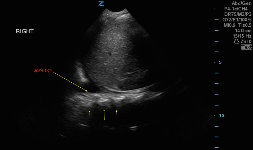
IOTW: Nephrotic syndrome with pleural effusion
This is an image of the RUQ, showing a pleural effusion. For orientation, the head is to the left of the screen and the feet are to the right of the screen. This is an example of pleural effusion. If the lung is full of air (normal) then you shouldn’t see the spine superior to […]
POCUS IOTW: Testicular Torsion
This is a 16 year old boy at UMC who had right testicular pain a few hours ago. The bedside US initially shows no flow and then he was manually detorsed. The second image shows that the patient’s right testicle subsequently had flow. As you can see, the color flow map on the left shows […]