
This post illustrates the difference between a simple renal cyst and hydronephrosis. The cyst is located within the cortex, is anechoic, well-circumscribed, does not branch out into the kidney, and is not connected to the ureter. In contrast, hydronephrosis is not well-circumscribed, it branches into the kidney, and is connected to the ureter.
Author: Mary Kate Claiborne
Mary Kate is from Augusta, Georgia. She attended Notre Dame for undergrad and then quickly returned to the warm weather in the South, attending medical school in Savannah, Georgia. She completed residency at University of Texas at Houston and PEM fellowship at Phoenix Children's Hospital. She is currently working as a PEM attending while also completing an ultrasound fellowship at CNMC. In her free time, Mary Kate enjoys running, tennis, cooking, spin class, coffee, and visiting family.
IOTW: Elbow effusion
This patient presented after injury to the elbow. POCUS was performed looking at the posterior elbow which identified an effusion (which in the setting of trauma is blood). Xray confirmed supracondylar type I fracture.
IOTW: Dilated Cardiomyopathy
This patient is a 9yo F with PMH of dilated cardiomypathy who presented to the UMC ED for fever and tachycardia with a family member who was influenza A+. Cardiac POCUS was performed. Patient was noted to have enlarged left ventricle with grossly decreased function. Left ventricular ejection fraction (LVEF) was estimated using E-point septal […]

IOTW: Knee effusion
A 16yo F presented for 1 week of right knee pain and 2 days of knee swelling. POCUS of the knee was performed which revealed a knee effusion. Knee effusions will present as fluid under the tendon. If fluid is visualized above the tendon, then other diagnoses such as cellulitis, abscess, hypoproteinemia, hematoma, etc should […]
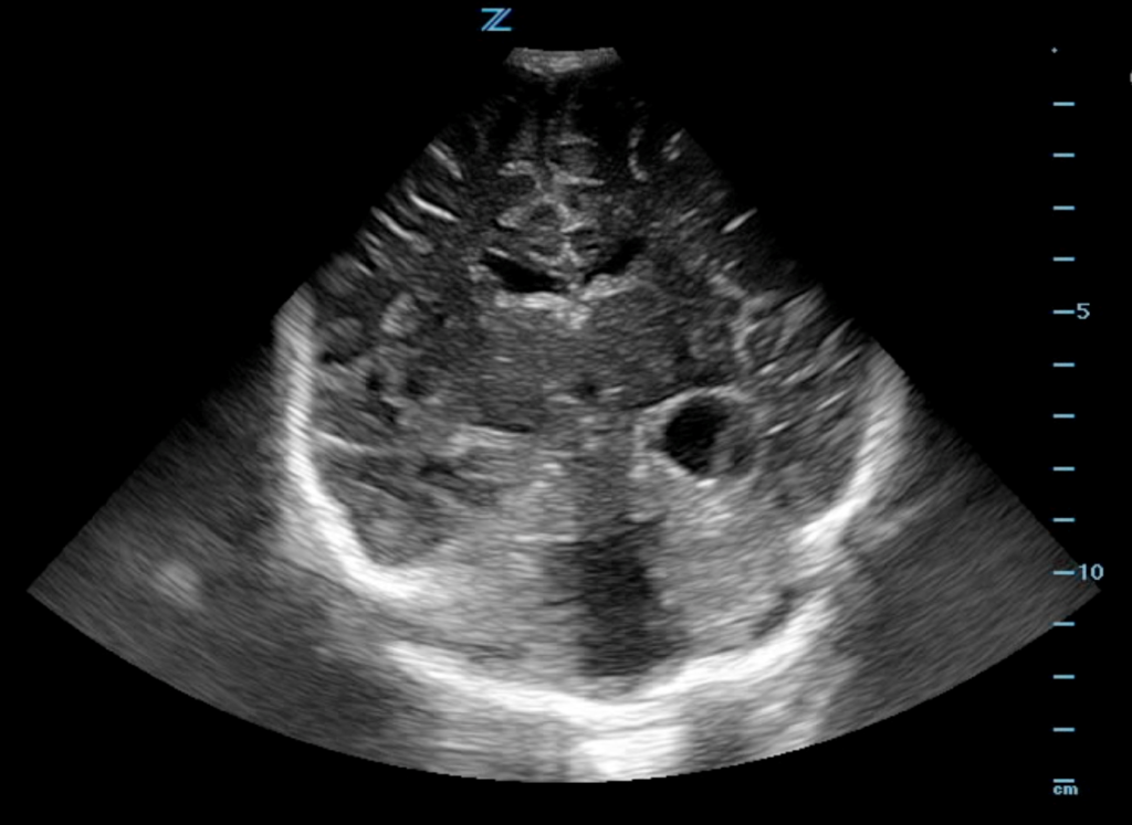
IOTW: Neo head ultrasound for Dandy-Walker malformation
Dandy-Walker syndrome is a congenital malformation that affects the development of the cerebellum. Typical features: the vermis is absent or hypoplastic, cystic enlargement of the fourth ventricle, enlarged posterior fossa. May be associated with hydrocephalus (this patient has normal sized ventricles). This three month old’s brain ultrasound demonstrates posterior fossa enlargement with cystic dilation of […]