
IOTW: Nephrolithiasis with Pelviectasis
Posted on: August 26, 2019, by : Simone Lawson
This week’s ultrasound shows the kidney of a 17 year old female presenting with left flank pain. She was diagnosed with a kidney stone two days prior but returned to the ED with worsening pain.
There is an echogenic renal stone with posterior shadowing as well as a dilated renal pelvis.
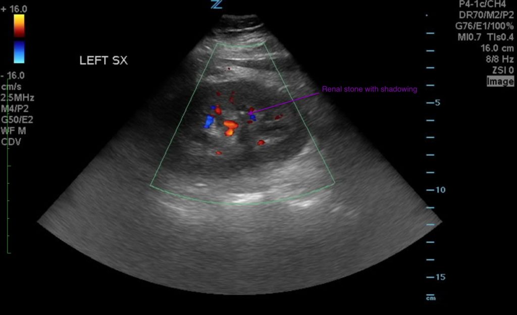

Although the renal vasculature, pelviectasis, and hydronephrosis will all appear as anechoic areas on ultrasound, color flow can help you differentiate between vascular and non-vascular findings.
Although not shown in the images above, “twinkling artifact” can be seen in the case of renal stones. It is the rapid alteration of color immediately behind a stationary echogenic object when using color doppler.
“The information in these cases has been changed to protect patient identity and confidentiality. The images are only provided for educational purposes and members agree not to download them, share them, or otherwise use them for any other purpose.”