
IOTW: Pericardial effusion
Posted on: September 17, 2019, by : Rosemary Thomas-Mohtat MD
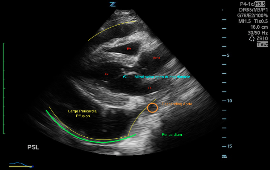
The image of the week is impressive. This teen had undifferentiated cardiomegaly on a chest Xray from an outside institution. POCUS was requested by the astute providers. You see 2 identical cardiac images of the parasteral long view (PSL) of the heart (one original, and the other annotated). From images of prenatal ultrasounds, even the layperson knows that black on ultrasound is fluid.There is a large black space surrounding the heart= Pericardial Effusion (931cc in this case).

How does one distinguish pericardial effusion from pleural effusion?
Pericardial effusion is above the descending aorta and often crosses the midline above the aorta. Pleural effusion is seen below/posterior to the descending aorta and does not cross the midline.

We’re taught that tamponade physiology is a clinical diagnosis (with only small % exhibiting Beck’s Triad of JVD, muffled heart sounds and hypotension), but FOCUS (focused cardiac ultrasound) is a good adjunct to evaluate RV collapse during diastole. In this image, you see the mitral valve fully open during diastole, and RV is relatively intact. The workup is still underway for the patient for the final diagnosis.
“The information in these cases has been changed to protect patient identity and confidentiality. The images are only provided for educational purposes and members agree not to download them, share them, or otherwise use them for any other purpose.”