
IOTW: Occipital Swelling
Posted on: June 17, 2019, by : Simone Lawson
This week’s image is a three year old with one week of occipital swelling.
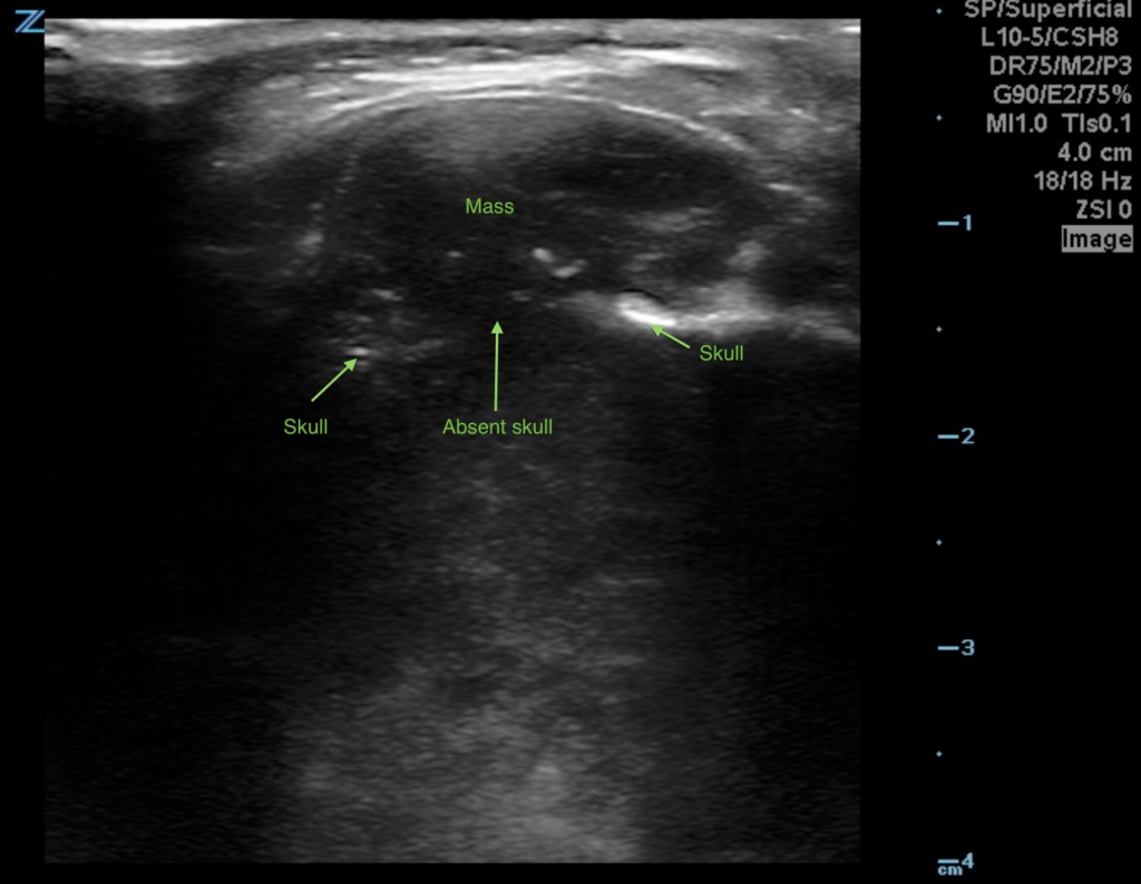
Beneath the mass, you will notice there is discontinuity of the skull representing erosion from what was determined to be a bony occipital tumor.
Remember that bone will show up as hyperechoic (white) on ultrasound.
When evaluating masses, always check for flow before presuming it is an abscess or lymph node. This mass did not have surrounding increased flow (on other images). You would expect stalk-like flow to a lymph node. An abscess would have absent flow within and increased flow surrounding it. A cyst would be hypo/anechoic without surrounding flow.
“The information in this case has been changed to protect patient identity and confidentiality. The images are only provided for educational purposes and members agree not to download them, share them, or otherwise use them for any other purpose.”