
This week’s image shows how artifacts can help you identify pathology on ultrasound. Drs. Batabyal and Zhao cared for a child with a wooden foreign body in her leg. Notice that the soft tissue appears normal and you cannot see the foreign body itself, but the shadowing present lets you know a foreign body is […]
Author: Carrie Ng
Carrie was born in San Francisco, but grew up between Hong Kong and Los Angeles. Although she loves the sun, she was excited to move away from home to Philadelphia for medical school, where she explored a new city, learned to appreciate all four seasons, and met her Canadian husband. She then moved to New York City and completed her Pediatrics residency at NYU and PEM fellowship at Columbia. During this time, Carrie developed a passion for bedside ultrasound and decided to join the CNMC faculty while pursuing an Emergency Ultrasound fellowship. In her spare time, Carrie loves traveling, listening to podcasts, and playing board games.
IOTW: Lung Ultrasound
Lung ultrasound can be useful in elucidating the cause of acute dyspnea. This week’s images are of a 2 year old female with respiratory distress and fever. In image 1 there is normally aerated lung viewed in the transverse plane as evidenced by the smooth white pleural line and A line artifacts. In image 2 […]
IOTW: Unilateral hydronephrosis
19 year-old-girl with right flank pain found to have mild unilateral hydronephrosis. You can differentiate renal vessels from the ureter by adding color doppler. The patient was also found to have a 5 mm urolithiasis at the UV junction on radiology US that is likely the etiology of the hydronephrosis and was discharged home with […]
IOTW: Ankle Fracture
Ultrasound can also be used to identify bony injury – notice the disruption of the cortex of the distal fibula of this teenager who jumped off a wall. Normally bone appears hyperechoic (bright white) with shadowing behind. Remember that growth plates will also appear as a disruption in the cortex, but this tends to be […]
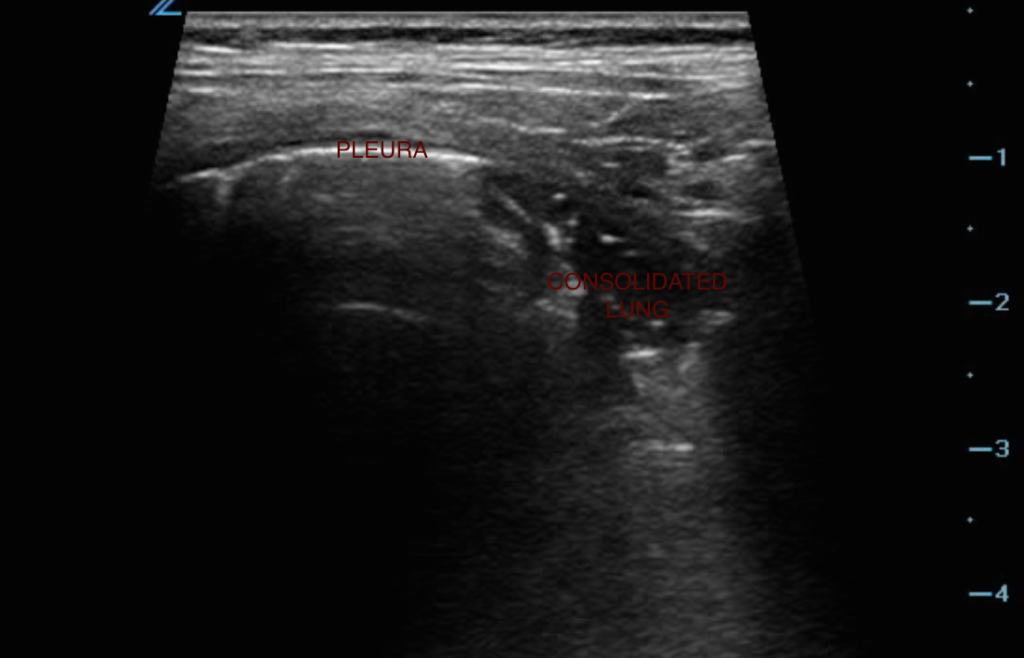
POCUS IOTW: Empyema
This is a teenage girl who presented with shortness of breath, right-sided shoulder pain and back pain. On ultrasound, she was found to have a large complex effusion with septations concerning for empyema. The phased-array transducer was used in order to have the depth to capture the fluid collection in the right chest. On CT […]