
IOTW: Lung Ultrasound
Posted on: May 16, 2019, by : Carrie Ng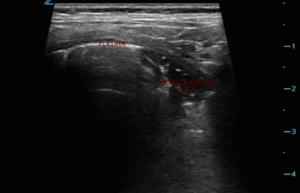
Lung ultrasound can be useful in elucidating the cause of acute dyspnea. This week’s images are of a 2 year old female with respiratory distress and fever.
In image 1 there is normally aerated lung viewed in the transverse plane as evidenced by the smooth white pleural line and A line artifacts.

In image 2 the white pleural line is interrupted by an irregularly shaped area concerning for consolidation

The sonographer, Dr. Lawson, appreciated this difference in the transverse plane and interrogated the area further by evaluating it in the longitudinal plane.
Within this view, the sonographer rotated the probe 90 degrees so that the ribs are out of view and the image is focused on the lung. In this image we are able to appreciate that the pleural line is interrupted by a shredded appearing border that almost appears as if a chunk has been taken from the lung, this is a consolidation.

This area can be compared to the 4th image of normally aerated lung without a consolidation in the longitudinal plane.

“The information in these cases has been changed to protect patient identity and confidentiality. The images are only provided for educational purposes and members agree not to download them, share them, or otherwise use them for any other purpose.”