
Hot Seat #221: Making The Breast Choices
Posted on: January 19, 2024, by : Brandon Ho
Case by Brandon Kappy MD, CNH PEM Fellow
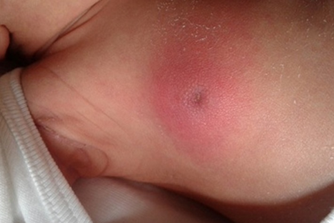
A 35-day-old female presents to the ED with what Mom describes as a “right-sided chest rash.” Mom states that she noticed the mild erythema around 2 days ago when she was breastfeeding the infant. Since then, Mom believes that it has become moderately more erythematous, though no enlargement or drainage. It may also be tender, as the infant is increasingly fussy when Mom touches the area. Otherwise, infant has felt warm at home, though no fevers recorded or other ill symptoms. No other rashes or contacts with a similar rash. Infant is breastfeeding well with a normal amount of wet diapers.
PMH: Infant was born full-term and has no other past medical history. All prenatal labs and screens normal. Normal vaginal delivery; received HepB and Vitamin K at birth. Discharged from hospital next day.
Vital Signs T 37.1 C, HR 140s, BP 82/52, RR 22, O2 100% RA
General Well-appearing infant, appropriate for age
HEENT Normocephalic, atraumatic. EOMI, no conjunctival erythema/injection. Oral mucosa moist. No pharyngeal erythema or exudate.
CV Regular rate and rhythm. Normal peripheral perfusion. Brisk cap refill.
Pulmonary Lungs clear to auscultation; non-labored respirations. Breath sounds equal bilaterally.
Abdomen/GU Soft, nontender, non-distended, normal bowel sounds. Normal GU exam.
Neuro No focal neurologic deficits observed. Developmentally normal.
Skin Unilateral R-sided erythema over the breast. No fluctuance palpated; mild induration of area without any pustular discharge or bleeding. Very scant clear nipple discharge when palpated with pressure. No crusting. Infant is crying during exam but area may also be tender on palpation. Normal L-sided breast w/o rashes. No other rashes noted on exam.
You obtain a CBC, CRP, Procalcitonin, Blood Culture, Urinalysis, and Urine Culture. You also obtain an ultrasound. Infant has remained well-appearing, afebrile, and breastfeeding normally during the workup. The labs and imaging result:
CBC WBC 15.2, Hb 12.2, PLT 234, ANC 4800
CRP 28 mg/L
PCT 0.2 ng/mL
UA Normal
Soft Tissue U/S Poorly defined hyperechoic right breast tissue with hyperemia and thickened subcutaneous tissue. No radiographic signs of abscess formation.
After discussion with a colleague, you elect to send swabs (on a scant amount of clear discharge) but not perform an LP.