
Hot Seat #81 Denouement: 15 mo M with URI symptoms and leukocytosis
Posted on: October 6, 2016, by : Evan Sherman
Jaclyn Kline, Children’s National Medical Center
The Case
15mo M F is referred from his PCP after he was found to have leukocytosis in the setting of URI sx. He had focal findings on lung exam, and a subsequent chest x-ray was abnormal.
Here’s How You Answered Our Questions:
Discussion:
Nearly everyone agreed that a chest x-ray was indicated in this toddler with leukocytosis and focality on his lung exam. Everyone at our in-person debrief also agreed that the x-ray was difficult to interpret. There was concern for tracheal deviation, although Jennifer reminded us that a larger degree of tracheal deviation is tolerated in young patients whose tracheal cartilage is still developing. In addition, this x-ray was a bit rotated, making assessment of tracheal deviation difficult. While the density in the right lung could have been due to the film being shot while the pt was exhaling, it made it difficult to rule out pneumonia or a mediastinal mass. There were relatively few votes for obtaining a chest CT in the polls, but the room seemed to be convinced that it was the right thing to do after we finished our discussion.
Comments from Dewesh and Pavan provided excellent explanations for their clinical decision making in this case.
Denouement:
The xray was discussed with the radiology resident on call, who read it as:
Dense opacification of the central hilar area with rightward bowing of the trachea. Prominent appearance of the cardiothymic silhouette. While findings could relate to expiratory film, difficult to exclude mediastinal mass and/or airspace disease. Recommend repeat radiograph for better inspiration, or CT.
The remainder of the labs also come back:
WBC – 20.2, 11 segmented neutrophils, 1 band, 43 lymphocytes, 19 monocytes, 1 eosinophil, 25 reactive lymphocytes
Hgb – 12, Hct – 32.8, Plt – 198
CMP – normal
Uric Acid – 4.7 (1.7-5.0), LDH – 293 (164-286)
A chest CT was obtained, which showed:
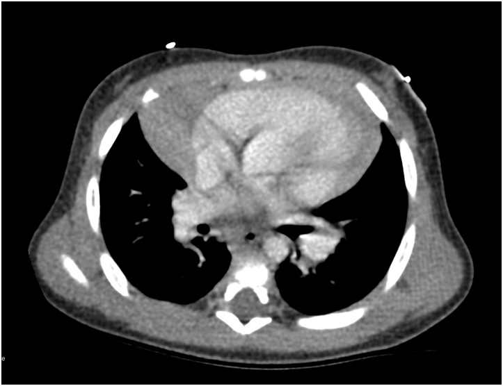
The final read of the CT was:
Normal-appearing thymus with no mediastinal mass. Bibasilar airspace opacities which may represent developing pneumonia versus focal atelectasis.
The child was ultimately diagnosed with viral bronchiolitis and an enlarged thymus. He was discharged home with continued supportive care and PCP followup.
The information in these cases has been changed to protect patient identity and confidentiality. The images are only provided for educational purposes and members agree not to download them, share them, or otherwise use them for any other purpose.