
Hot Seat Case #104: 10 week old female with emesis
Posted on: December 4, 2017, by : Hilary Ong
Hilary Ong, MD Children’s National Medical Center
with Lenore Jarvis, MD Children’s National Medical Center
10 week old ex-FT female with no significant PMH presented with fever Tmax 101 and vomiting for 3 days and 1 day of intermittent increased work of breathing. She had not had any nasal congestion or cough recently, but siblings at home had URI symptoms in the past week. One day prior to arrival, she had 4 NBNB emesis, each after a feed. Her last emesis was 3hrs prior to presentation. Her abdomen per mom was very distended.
ROS: Decreased appetite. Breastfeeding and taking formula frequently with less volume each feed. Has had normal wet diapers. Fussy and difficult to console despite Tylenol PRN for fever and fussiness. No diarrhea. No rash.
Birth History: FT, NSVD, no complications during delivery
PMH: none
PSH: none
Vaccinations: up to date
PE: T 38.5 HR 190 RR54 BP 115/79 SaO2 99%
General: Alert. well appearing. fussy, difficult to console.
Skin: Warm. dry. intact. no pallor. no rash. normal for ethnicity.
Head: Normocephalic. atraumatic. anterior fontanelle soft and flat.
Neck: no lymphadenopathy.
Eye: Normal conjunctiva
Ears, nose, mouth and throat: Tympanic membranes clear. Oral mucosa moist. No pharyngeal erythema or exudate.
Cardiovascular: No murmur. No gallop. + tachycardia, regular rhythm. 2+ femoral pulses.
Respiratory: Lungs are clear to auscultation. Symmetrical chest wall expansion. Tachypneic with mild subcostal retractions.
Gastrointestinal: Soft. Normal bowel sounds. Abdomen distended . No hepatosplenomegaly.
Musculoskeletal: Moves all extremities
Neurologic: Alert, non focal neurologic examination
She was given acetaminophen for fever/pain and a 20 cc/kg NS bolus for tachycardia.
Labs:
CBC: 17k WBC, 9.9 Hb, 30.2 Hct, 306k Plt
28% Neutrophils 4% Bands 53% Lymphocytes 4% Reactive lymphocytes
Blood culture: negative gram stain
UA: negative LE, negative Nitrite, 2RBC, 1 WBC,
Urine culture: negative gram stain
The patient continued to have intermittent fussiness, tachypnea with a persistently distended abdomen.
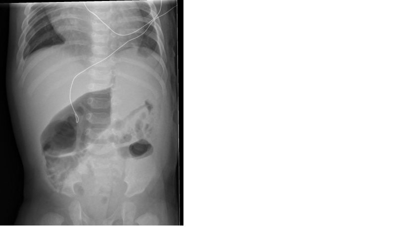
The KUB is now available and is as shown:
The information in these cases has been changed to protect patient identity and confidentiality. The images are only provided for educational purposes and members agree not to download them, share them, or otherwise use them for any other purpose.
Following on the heels of Rachel’s case last week, another vomiting infant. I know for the purposes of Hot Seat, sometimes there are omissions.
But I want to know more.
We’ve been focusing on EHR documentation recently. Sometimes, pre-fab templates can cause unintentional omission of necessary information. Wordy, I know. Remember our morning at Hopkins and the definition of CLARITY. Simply: you need to write a chart as though you are writing it for yourself in about two years time.
For example, the febrile neonate with vomiting: If I examined the infant and did not feel the urge to proceed with an LP, I would want to be able to document (besides that the infant appeared well), that the fontanelle was not bulging, neck was supple, tone was normal, red reflexes. Jah?
Back to the vomiting, though, and age based differentials. I’ll let folks chime in before I say anything, it was great to see fellows contribute the last time.
Super brief note: KUB is a single view of a supine patient. With vomiting or any symptom concerning for obstruction, 2-views are required: the KUB plus an upright, a X-table or a left lateral decubitus view.
Jennifer, good point. My wording is indeed inaccurate. For this case presentation, and for purpose of clarifying the last question, let’s say the patient did initially get a 2-view abdominal XR AP/PA and upright.
It’s hard for me to fully read that KUB, but my preliminary read would be concerning for an obstruction as it does not appear that gas is tracking to the rectum. Most cases of malrotation present before the age of one and the presentations don’t have to be bilious emesis. From an ED perspective, with a distended abdomen (appreciated by mother and the ED doc), I think our job is to rule out malrotation/volvulus leading to peritonitis/perforation. It’s hard for me to justify doing something else (spinal tap, watching on abx), when it seems the clinical exam and history is pointing to the belly… Although if the clinical status is worsening while waiting for the UGI, I would go ahead with fluid boluses and broad spectrum GI antibiotic coverage.
I also think this kids heart looks big! Perhaps it’s just a single supine view and that’s causing the problem and the patient doesn’t look fluid overloaded, but poor perfusion from myocarditis or congenital heart disease can cause gut ischemia. Just a thought, and would get dedicated chest films as well.
I agree with Jeremy – it’s really hard to tell what’s going on with the AXR. I think it’s small bowel dilation that we see – are those mucosal folds of the distended area crossing the full width of the bowel? I’d definitely go with the upper GI as the next study. I think I would only consider an LP and antibiotics if the patient looked toxic and the tachycardia to 190 at triage didn’t resolve.