By Sonny Tat, Children’s National
with Shilpa Patel, Children’s National
The Case
 This is a case of a 3 year-old healthy female with abdominal pain and bloody stools who presents with blood at the anus and anemia. The challenge in this case is how to approach this patient with an ultrasound negative for intussusception and how to address access issues (a common problem). For a complete case presentation, please click here.
Here’s How You Answered Our Questions
What is your next diagnostic step in the ED?
Less than 3 years PEM experience (fellows, PAs/NPs, pediatricians) n=9
More than 3 years PEM experience (PEM attendings, pediatricians) n=7
Attendings and fellows agree that the next diagnostic step would be an abdominal x-ray. Interestingly, 22% of fellows would get an abdominal CT, while none of the attendings thought a CT the next step. Approximately 14% of attendings thought a barium enema might be beneficial, while none of the fellows chose that imaging modality.
You have been unable to establish IV access despite multiple attempts. What is your access plan?
Less than 3 years PEM experience (fellows, PAs/NPs, pediatricians) n=8
More than 3 years PEM experience (PEM attendings, pediatricians) n=7
The majority of providers would continue to attempt a non-EJ IV, followed by an EJ IV. This patient is sick, but stably so at the moment. She definitely needs access, but the majority of providers felt they could take the time to get an IV. Only 12.5% of fellows (and no attendings) felt an IO was necessary. No one wanted PICU admission for a central line. At this time, the patient doesn’t truly need ICU care.
Clinical Reasoning
Dave Mathison and Shilpa Patel (Hot Seat Attending) gave excellent responses to this case. For their complete comments, click here.
“it’s super, super important to differentiate painless versus painful rectal bleeding. and make sure it’s actually blood.”
“typically, PAINLESS rectal bleeding is either juvenile polyp or a Meckels. period. in this patient who has a drop in Hb with normal indices yet is well appearing….sounds like a Meckel’s. and if this were the case, i would admit for a meckel’s scan since the patient is minimally tachycardic with a low Hb and question of active bleeding. always good idea to get a PT and PTT to look for other coagulopathies, although this is not a presentation of acute hemophilia. and never a bad idea to put in an EJ, although probably not necessary if BP is normal and kid looks fine.”
“now PAINFUL rectal bleeding… think about infectious colitis versus inflammatory bowel disease versus vasculitis etc. so send the culture, ESR/CRP. If inflammatory markers are negative, this points more towards anatomic (meckels, polyp). If positive this supports one of these inflammatory or infectious conditions… I do like to get an abdominal XR, especially in a 3yo where ingestion of foreign material is possible… rectal obstruction can cause gaseous distention (pain) and bleeding from local trauma. same goes with constipation, although pretty HARD (no pun intended) to get your Hb down so much with constipation/tears.”
-Dave Mathison, Children’s National
From the Hot Seat
by Shilpa Patel, Children’s National Medical Center
Although lower GI bleeding in children may be indicative of a serious illness, usually it is not. Our very own Stephen Teach published a retrospective review of 109 cases (?his fellow’s project – pretty awesome) in which only four had life threatening diagnoses (https://www.dropbox.com/s/b53127f2haoqj43/1-s2.0-S0196064494703507-main.pdf) – three cases of intussusception and a single case of Meckel’s diverticulum.
The first step in the approach to GI bleed is to determine if it is truly blood. In our case, given the low hemoglobin and gross blood at the anus on exam, we should assume it is blood. The second step is to try to distinguish between upper and lower GI bleeding. Classic teaching is that bright red blood in/on stool is usually lower, however in an infant (short intestinal transit time), bright red blood could be from a massive upper GI bleed and quickly lead to hemodynamic compromise). Whereas melena, or dark blood is classically more proximal (this could be blood that is swallowed from the nose/ingested). Red stool (though it is dark) with gross blood at the anus in our patient is probably lower GI.
I’m going to use SPI(T) (which Sonny introduced to us a few weeks ago as a teaching technique in the ED) to organize my DDx and will try to limit it to younger children since our patient is 3 years old.
S Serious
P Probable
I Interesting
T Treatable
DDX:
S: Serious
Intussusception – Though this has already been ruled out (US has a negative predictive value of 99.5% with a good sonographer), our patient’s presentation, a 3 yo with intermittent severe abdominal pain sounds concerning. Blood in stool is a late finding in intussusception. The currant jelly stool is associated with necrotic bowel and I would expect the patient to appear more ill. Thinking creatively, the bloody stools in our scenario could be related to a lead point for intussusception (a bleeding polyp or diverticulum) causing the bleeding and the intermittent abdominal pain AND the telescoping could be intermittent (giving us a negative ultrasound when the child is comfortable).
Meckel’s diverticulum – usually described as “painless” bleeding (just blood and no stool), though our child reports abdominal pain. It can also be associated with complications (obstruction or perforation) and majority of the patients are younger than 2 years old. As Meckel’s can lead to rapid painless blood loss and hemodynamic compromise, ensuring that the patient is hemodynamically stable is very important. Our patient is anemic and should be watched. At this point, I don’t think I would get a Meckel scan on our patient.
Underlying coagulopathy – normal INR is reassuring along with normal platelets. HUS can present with GI bleed but is unlikely given normal platelets and normal Creatinine.
Volvulus (w/Malro) – would expect the child to be sicker with an abnormal aXR, or have bilious emesis (though she did have emesis x 2 earlier in the day). If she had bilious emesis, would consider an AXR.
P: Probable
Infectious colitis – Our patient has crampy abdominal pain, it is associated with multiple stools and had vomiting earlier -fits the bill. We should ask a travel/exposure history, send a second set of cultures and follow up on the stool cultures sent by the primary care doctor. Would also be good to know if she was on antibiotics (C. Diff colitis) Something that goes against this is the lack of fever and perhaps a normal wbc….
Mnemonic for infections that cause bloody stools: YEECCSS!
Y – Yersinia
E – Ecoli
E -Entamoeba histolytica
C – Cdiff
C- Campylobacter
S- Shigella
S – Salmonella
HSP – Our patient had a URI two weeks ago, has abdominal pain with LGI bleed, is the right age (majority of cases present between 3-5) and has normal renal function labs (at least for now, as 50% of HSP eventually have renal involvement). But…our patient does not have rash…which is fairly common…however I don’t think lack of rash rules it out…we need a scope.
IBD – 3 is a little young for onset of IBD though not impossible; the child has crampy abdominal pain with multiple blood stools (and classic relief of pain with bloody stool); rectal UC (more than Crohns) presents with LGI bleeding like this. The borderline low MCV of 79, microcytic anemia, could be suggestive of chronic GI blood loss or could be some underlying iron deficiency anemia. Also, there are no skin tags on rectal exam.
Juvenile (Rectal) Polyps – usually presents with bright red blood and is often painless. Can be a lead point for intussusception or volvulus so therefore could have abdominal pain with presentation. These can be anywhere in the GI tract (upper or lower) and hence it could be dark red blood if a bit more proximal. A rectal exam may allow the palpation of a rectal polyp (and these can pop out – often confused with rectal prolapse – I recently saw a case).
Gastric ulcer – could cause pain and bleeding; would be worried about perforation with significant bleed, but our patient looks well. Usually eating makes the pain worse (compared to better after eating with a duodenal ulcer). Would be good to know if she was on NSAIDs for any other reason.
Anal fissures – common but not likely in our patient given dark red stools and anemia…usually more benign
Allergic colitis: Eosinophillic proctitis can cause lower GI bleed (or upper GI if severe eosinophillic/allergic esophagitis)…would expect this to be more chronic
I: Interesting
Portal hypertension: There was a case report of a child with undiagnosed idiopathic portal hypertension presenting with an esophageal varices and bleed – highly unusual but interesting – though unlikely in our well appearing patient with intermittent bleeding and no HSM. Variceal bleeds are more rapid and quickly destabilize.
Gastrointestinal Duplication – can ulcerate, perforate or form fistulas, causing lower GI bleed. Very uncommon…we need a scope to diagnose this. I guess this could cause pain if ischemic.
Tumors/Malignant polyps and Vascular Malformations …very rare, again would need a scope
T: Treatable
Of the list above, many are treatable. I would use this question….to assist me in my management.
Management
For our patient, I agree with the testing that has already been done. I would follow up with the PCP regarding the stool cultures and probably send another set. I would send a type and cross, make sure I have solid access and admit the patient for observation given the low hemoglobin and ongoing blood loss. If IV access was difficult I would consider an EJ. Also, I would discuss this patient with the PICU as GI bleed can lead to rapid hemodynamic instability; and with GI to consider scoping.
The Denouement
by Sonny Tat, Children’s National
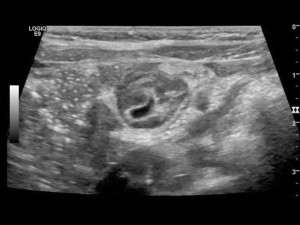
The patient continued to have intermittent pain so underwent a repeat abdominal ultrasound four hours later to evaluate the possibility of intermittent intussusception. This ultrasound demonstrated a thick walled cystic structure below the umbilicus with mild hyperemia without signs of intussusception. There was point tenderness at this location on ultrasound exam.
This patient remained hemodynamically stable and was admitted to the surgical service for further observation and diagnostic evaluation.
The patient had a nuclear medicine study showing radiotracer uptake below the umbilicus (figure 2). She then underwent a diagnostic laprascopy that was converted to an open resection of a Meckel’s Diverticulum. The patient had an uncomplicated post-operative course. Her final pathology showed heterotopic gastric tissue, consistent with Meckel’s Diverticulum.
Please answer the two quick questions below. The data helps inform our future case design. Thanks for participating.
Like the Hot Seat? Don’t miss a case. Subscribe for email updates of new posts by adding subscribing in the field at the top of this page or by clicking here.
The Hot Seat is a recurrent online case series aimed at facilitating asynchronous sharing of PEM knowledge and experience. Faculty are placed on The Hot Seat and publish their opinions without knowing the case or its outcomes. Cases are based on real cases and written by PEM fellows at Children’s National in DC, Johns Hopkins in MD, and INOVA Fairfax in VA to highlight teaching points. Emily Willner is the faculty advisor for the Hot Seat.

 This is a case of a 3 year-old healthy female with abdominal pain and bloody stools who presents with blood at the anus and anemia. The challenge in this case is how to approach this patient with an ultrasound negative for intussusception and how to address access issues (a common problem). For a complete case presentation, please click here.