IOTW: Elbow effusion
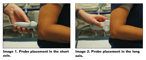
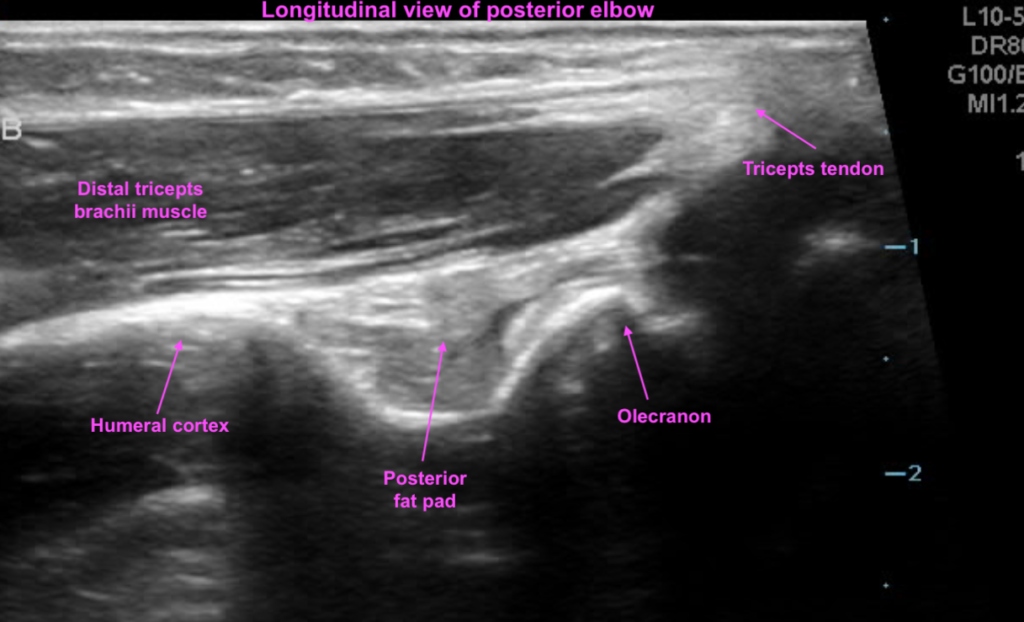
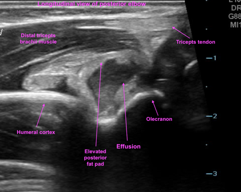
Posted on: February 28, 2020, by :

This patient presented after injury to the elbow. POCUS was performed looking at the posterior elbow which identified an effusion (which in the setting of trauma is blood). Xray confirmed supracondylar type I fracture.

This image demonstrates probe placement. Patient holds arm in flexed position. For the short axis view, hold the probe in the transverse position. For the long axis view, hold the probe in the longitudinal position.
This is a view of the elbow in the longitudinal plane. The posterior fat pad rests within the olecranon fossa.
This is a view of the elbow in the transverse plane. The posterior fat pad rests within the olecranon fossa.
This is a view of the elbow in the longitudinal plane. The posterior fat pad is elevated and there is an effusion within the olecranon fossa.
This is a view of the elbow in the transverse plane. The posterior fat pad is elevated and there is an effusion within the olecranon fossa.

Leave a Reply

Your email address will not be published. Required fields are marked *